Posterior Triangle of the Neck
Boundaries
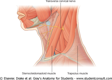
Trapezius muscle
Sternocleidomastoid
Middle third of tje clavicle
Roof:
Investing layer of deep cervical fascia
Floor:
Splenius
Levator scapulae
Scalenus medius
Scalenus anterior and first digitation of Serratus anterior may contribute to the floor.
At the apex Semispinalis might appear
Contents
Artery
Subclavian artery
Nerves
Trunks of the brachial plexus
Branches of cervical plexus
The above-mentioned contents are covered by prevertebral fascia
Note that a surgoen wont cause injury to these structures if he does not breach the overlying prevertebral fascia.
Accessory nerve (CNXI)
Lymph nodes
Occipital (2 or 3 in number)
Supraclavicular
Muscle
Inferior belly of omohyoid