Monday, 22 February 2016
Triangles of the Neck
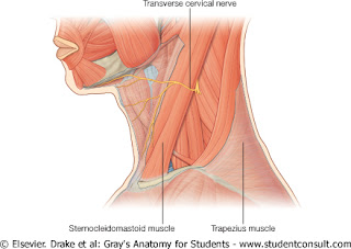
Posterior Triangle of the Neck
Boundaries
Trapezius muscle
Sternocleidomastoid
Middle third of tje clavicle
Roof:
Investing layer of deep cervical fascia
Floor:
Splenius
Levator scapulae
Scalenus medius
Scalenus anterior and first digitation of Serratus anterior may contribute to the floor.
At the apex Semispinalis might appear
Contents
Artery
Subclavian artery
Nerves
Trunks of the brachial plexus
Branches of cervical plexus
The above-mentioned contents are covered by prevertebral fascia
Note that a surgoen wont cause injury to these structures if he does not breach the overlying prevertebral fascia.
Accessory nerve (CNXI)
Lymph nodes
Occipital (2 or 3 in number)
Supraclavicular
Muscle
Inferior belly of omohyoid
Wednesday, 3 February 2016
Deep Fascia of the Neck
Surrounds the neck like a collar and consists of four parts:1- Investing Layer
Splits around Trapezius and Sternocleidomastoid muscle
Attachments:
Anteriorly, to the hyoid bone.
Posteriorly, it blends with Ligamentum nuchae.
Superiorly, to the lower border of the mandible, mastoid process, superior nuchal line and external occipital protuberance.
Inferiorly, to acromion, spine of the scapula, clavicle and manubrium of the sternum.
Between the angle of Mandible and styloid process the fascia splits to enclose the parotod gland. The posterlayer forms the stylomandiblar ligament
2- Prevertebral
Firm tough memebrane that lies in front of the prevertebral muscles
extends laterally in front of the scalenus anterior, medius and levtor scapulae
Fadesbehind Trapezius muscle.
3- Pretracheal
Lies deep to the infrahyoid muscles
Splites to enclose the thyroid gland and attaches to the second, third, and fourth tracheal rings.
4- Carotid Sheath
Surrounds the common carotid artery, internal carotid artery, internal jugular vein and vagus nerve. Attaches superiorly to the base of the skull and inferiorly blends with the adventitia of the aortic arch.
Tuesday, 2 February 2016
General topography
The hard palate lies at the level of C1 (anterior arch of atlas vertebra).The lower border of mandible lies between C2 and C3 vertebrae.
The pharynx extends from the base of the skull to the level of the cricoid cartilage (C6).
The hyoid bone lies at the level of C3 and is connected to the mandible by mylohyoid muscle.
In front of the lower phaynx and oesophagus lie the larynx and trachea.
Extensor musculature lies behind the vertebrae and supplied by dorsal rami of cervical nerves.
Flexor musculature lies anterior to the vertebrae and behind the pharynx and supplied by ventral rami of spinal nerves.
On each side of the pharynx is the carotid sheath containing :
The common carotid artery, the internal carotid artery and the internal jugular vein with cervical sympathetic trunk lying posteriorly.
The investing layer of deep cervical fascia surrounds the neck like a collar and attaches to the base of the skull superiorly and the clavicle and root of te neck inferiorly.
Subscribe to:
Comments (Atom)


